Using 3D-printed models of patients’ organs to diagnose urology conditions.
Ricky Crowe didn’t realize how big the tumor on his kidney was until he was in the recovery room.
His surgeon, Dr. Chris Keel, walked in with a three-dimensional, full-size model and Crowe saw for the first time the growth winding around his kidney.
“I’d seen the pictures, but I just didn’t understand it until he showed me that,” Crowe said. “It helped a lot.”
More patients at Erlanger hospital will have Crowe’s experience as a result of a contract the hospital signed Wednesday with 3D Ops, a local tech startup that hopes to launch a nationwide network of 3-D printing centers to give surgeons and patients a real-life look at damaged organs before starting surgery.
What’s key is that the model Keel used was not a normal kidney, but rather, a model of his patient’s actual kidney — with all of its abnormalities.
“It is Ricky’s kidney,” said Keith Campbell, president of 3D Ops. “You see the real thing.”
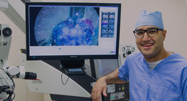
That can be a big help for a surgeon. Keel wanted the arteries to be printed in blue and the kidney in clear plastic, so he could see how to avoid slicing into the arteries when he cut out the tumor.
“There are some benefits in being able to see it in three dimensions and having a game plan when you get in there,” he said.
While he said he could have performed Crowe’s surgery without the aid of the model, it made it easier for him to remove the tumor without taking out the kidney as well.
Campbell said the idea for the process came three years ago, when he was talking with Dr. David Seaberg, dean of the University of Tennessee College of Medicine Chattanooga.
“We were looking at what you could do with 3-D printing and he said, ‘You know, we have all sorts of models of what is right. But we don’t have models of what is wrong,'” Campbell said.
The Erlanger contract is a major step forward in building what Campbell hopes will be a network of several dozen 3-D printing centers, serving hundreds of hospitals. 3D Ops’ goal is to turn around the models in 24 hours or less. The company has patents, Campbell said, on the software that converts images from CT scans or MRIs — the expensive machines hospitals use to capture an image of the insides of the body — into a format that can be used for 3-D printing.
The Erlanger deal also gives 3D Ops the opportunity to work with the hospital imaging staff to figure out the proper settings, or protocols, to use for each machine and each type of operation.
And it will give the company a chance to work with other surgeons to figure out more ways to use the models.
Keel believes the modeling would be particularly helpful for reconstructive surgery, where a doctor may need to bend pieces of metal around bones in order to reshape a face. If the metal could be shaped in advance using the physical model, he said, it could dramatically shorten the length of the operation. Campbell is also optimistic about future improvements in 3-D printing.
The technology has been around since the 1980s, but has made significant improvements in the past few years in terms of the materials that can be used, the level of detail that can be printed, printing speed and cost. The process works much like a traditional printer, with a print head moving across a space and spraying a layer of liquefied material. But instead of ink, metals such as copper or titanium can be used or various forms of plastic or even food products. Each pass of the printing head adds a new layer and over time those layers add up to form the 3-D model.
Many large teaching hospitals, where surgeons are trained, may be able to afford their own 3-D printing equipment, Campbell said.
But that still leaves more than 5,000 hospitals across the U.S. which are unlikely to buy their own equipment, given its complexity and how fast the technology is changing.
Campbell is not shy about his goals. In five years, 3D Ops hopes to have more than 300 hospitals in its network, printing an average of 11 models per hospital each month, at an average cost of $400 per model, plus a monthly subscription fee for each hospital.
“By the fifth year, we’re a $49 million company in revenue, $20 million in profit,” Campbell said.
Keel, a urologist who has been performing advanced kidney surgery for several years now, even foresees a time when a surgeon would use a detailed 3-D printed model of a damaged organ to work out exactly how to perform a surgery. Then the surgeon could upload those instructions to a robotic surgeon to perform the surgery on a patient located hundreds or thousand of miles away.
Read the original article here.